


$5,843.19
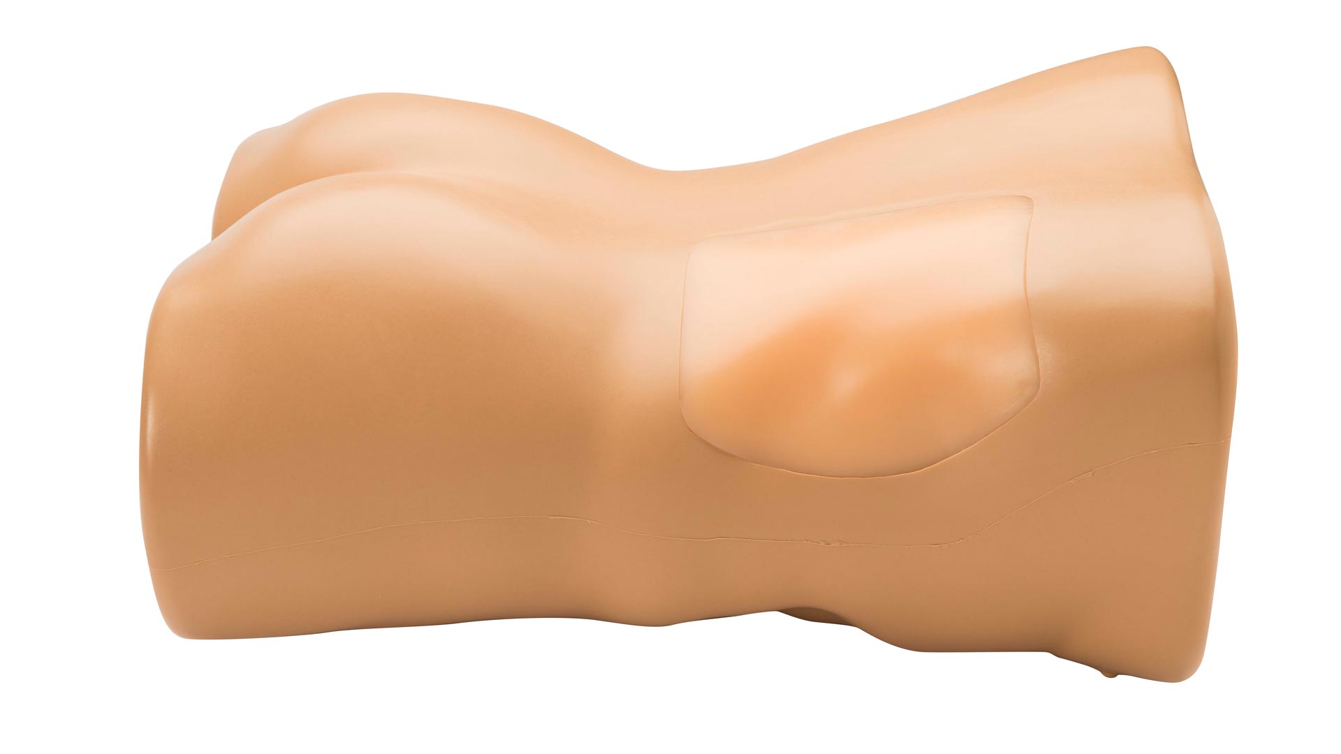
Elevate ultrasound-guided renal biopsy skills with high anatomical and procedural realism
Right kidney with internal detail for real-time sonographic targeting
Supports both core biopsy and needle aspiration techniques
Self-healing tissue allows for repeated needle access without degrading image quality
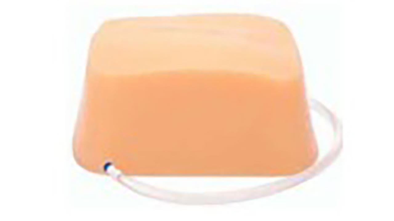
Removable kidney insert extends training life and reduces replacement costs
Train learners to identify the renal cortex, medulla, major and minor calyces, and surrounding tissue in real time using 2D ultrasound imaging.
Identify sonographic landmarks such as the right lobe of the liver, small bowel, and transverse colon within a lifelike abdominal cavity.
Practice real-time needle guidance into fluid pockets, reinforcing probe handling, needle visualization, and procedural confidence.
Perform full ultrasound-guided renal biopsies using 18–21 gauge needles. Learners can remove core samples or perform needle aspiration with realistic resistance and feedback.
Includes ribs and soft tissue, allowing learners to use ultrasound to avoid bone structures while positioning the transducer and guiding the needle safely.
Use the model repeatedly for needle aspiration thanks to its patented self-healing tissue. For core biopsy, the kidney module is easily rotated or replaced after 30 tissue samples.
Anatomy:
Dimensions:
Standard Coverage: 1-Year Manufacturer’s Warranty included
Develop the essential skills required to perform safe, accurate ultrasound-guided renal biopsies with this high-fidelity simulation model. Designed for clinicians training in nephrology, interventional radiology, emergency medicine, and ultrasound education, this model replicates the sonographic experience of performing percutaneous renal biopsy on an adult male patient.
The model includes a detailed, anatomically correct right kidney embedded in an adult-sized torso. Learners can use real-time 2D ultrasound imaging to identify key anatomical structures such as the renal cortex, medulla, and calyces, and practice navigating around the ribs to reach a safe biopsy site. Compatible with your own imaging system and transducers, it allows for full procedural practice including transducer handling, image interpretation, and needle placement.
The embedded kidney supports both core needle biopsy and needle aspiration techniques. The self-healing tissue offers extended utility for aspiration procedures, while the replaceable kidney insert can accommodate up to 30 core samples before being rotated or replaced—giving you a longer product life and more training per unit.