


$10,255.37
Realistic pelvic pathology for transvaginal ultrasound training using your own imaging system
Multiple fibroids, cysts, and masses with detailed female pelvic anatomy
Supports transducer insertion, manipulation, and pathology recognition
Durable, self-healing tissue with lifelike imaging across 2D, 3D, and 4D systems
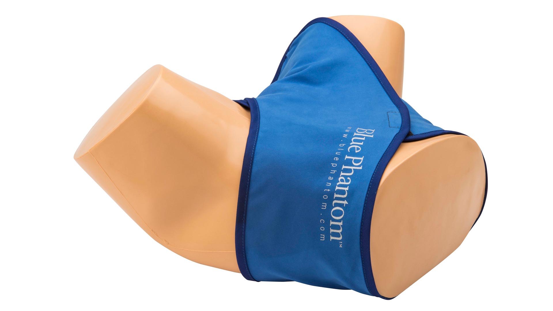
Includes privacy skirt for simulation-based training environments
Includes endovaginal and cervical canal, uterus with prominent endometrium, left and right ovaries, bladder with bladder wall, bowel and colon, broad ligaments, and accessory structures for full anatomical context.
Train learners to identify and measure uterine fibroids of various sizes, ovarian cysts, a large ovarian mass or abscess, ovarian follicles, and free fluid in the cul-de-sac—mirroring a variety of patient presentations.
Tissue composition replicates the acoustic characteristics of real human tissue for accurate sonographic imaging across 2D, 3D, and 4D platforms. Use your own endovaginal transducers (5.0–12 MHz recommended) for optimal realism
Simulates the tactile feel of performing an actual transvaginal ultrasound, allowing learners to practice correct insertion technique and internal probe navigation.
Self-healing tissue withstands repeated training use without the need for replacement parts. Easy to clean and maintain between sessions.
Includes a privacy skirt for realistic simulation settings and a soft storage case with foam block for easy transport and protection.
Anatomy:
Dimensions:
Included Accessories:
Standard Coverage: 1-Year Manufacturer’s Warranty included
Usage Requirements: For optimal performance and realistic simulation, use with an appropriate endovaginal ultrasound transducer (5.0–12 MHz recommended).
Developed for learners seeking hands-on experience in performing endovaginal ultrasound exams, this training model offers a highly realistic platform for building competency in image acquisition, transducer handling, and pelvic pathology interpretation.
The model presents a comprehensive range of anatomical structures and pathologic conditions including uterine fibroids, ovarian cysts, free fluid, and a large ovarian mass—providing varied learning scenarios for both basic and advanced gynecologic ultrasound applications. Learners can practice real-time transducer manipulation, identify pelvic anatomy, and utilize their own ultrasound systems to simulate live clinical imaging conditions.
Ideal for learners in obstetrics, gynecology, fertility medicine, emergency care, radiology, and medical education, this model supports skill development in transducer insertion, internal anatomy navigation, and use of ultrasound calculation tools for measuring cysts, masses, and follicles.