


Train on B-mode and elastography ultrasound imaging with lifelike tissue elasticity and a variety of target lesions
Contains soft, stiff, and isoelastic lesions with variable sonographic appearance
Supports elastography image acquisition and ultrasound-guided biopsy
Self-healing, maintenance-free SimulexUS™ tissue
Masses range from 6 mm to 11 mm in central breast and Tail of Spence
Practice identifying and targeting soft, stiff, and isoelastic lesions with different Young’s Modulus values—ideal for mastering elastogram interpretation and transducer pressure control.
Train using lesions that range from hypoechoic to hyperechoic and simulate the complexity of real patient exams.
Develop and refine ultrasound-guided biopsy skills using 18–21 gauge needles; fluid injections confirm accurate needle placement.
Self-healing SimulexUS™ tissue allows for repeated use without image degradation—a cost-effective solution for simulation centers and training programs.
Synthetic tissue won’t dehydrate or require special storage, ensuring long-term usability with minimal upkeep.
From novice sonographers to seasoned radiologists, users can progressively train on lesions of increasing difficulty in both size and location.
Anatomy:
Dimensions:
Included Accessories:
Standard Coverage: 1-Year Manufacturer’s Warranty included
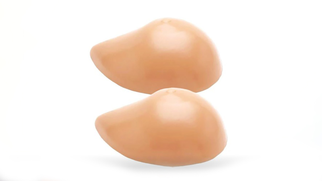
The Biopsy and Elastography Ultrasound Training Model from Blue Phantom provides a comprehensive, hands-on training platform for clinicians developing proficiency in both B-mode and elastography ultrasound imaging, as well as ultrasound-guided fine needle biopsy.
Crafted with Blue Phantom’s patented SimulexUS™ tissue, this model mimics the tactile feedback and imaging characteristics of real human tissue. It includes 12 lesions of varying viscoelastic properties—ranging from soft to stiff to isoelastic—and offers a wide range of echotextures, including hypoechoic, isoechoic, and hyperechoic masses. This variability makes the model ideal for practicing lesion identification, transducer pressure control, and guided interventions across a realistic clinical spectrum.
Lesions range in size from 6 mm to 11 mm and are distributed through both central breast tissue and the Tail of Spence, supporting skill development across different anatomical regions. Users can also inject fluid to confirm needle placement—fluid is automatically expelled for quick resetting between sessions.
The model is compatible with all major ultrasound systems and transducers (linear array, 7.5–15 MHz recommended), making it an excellent choice for simulation labs, clinical training programs, and product demos.