
$2,931.45
Master ultrasound-guided fine needle biopsy techniques with high-fidelity thyroid anatomy
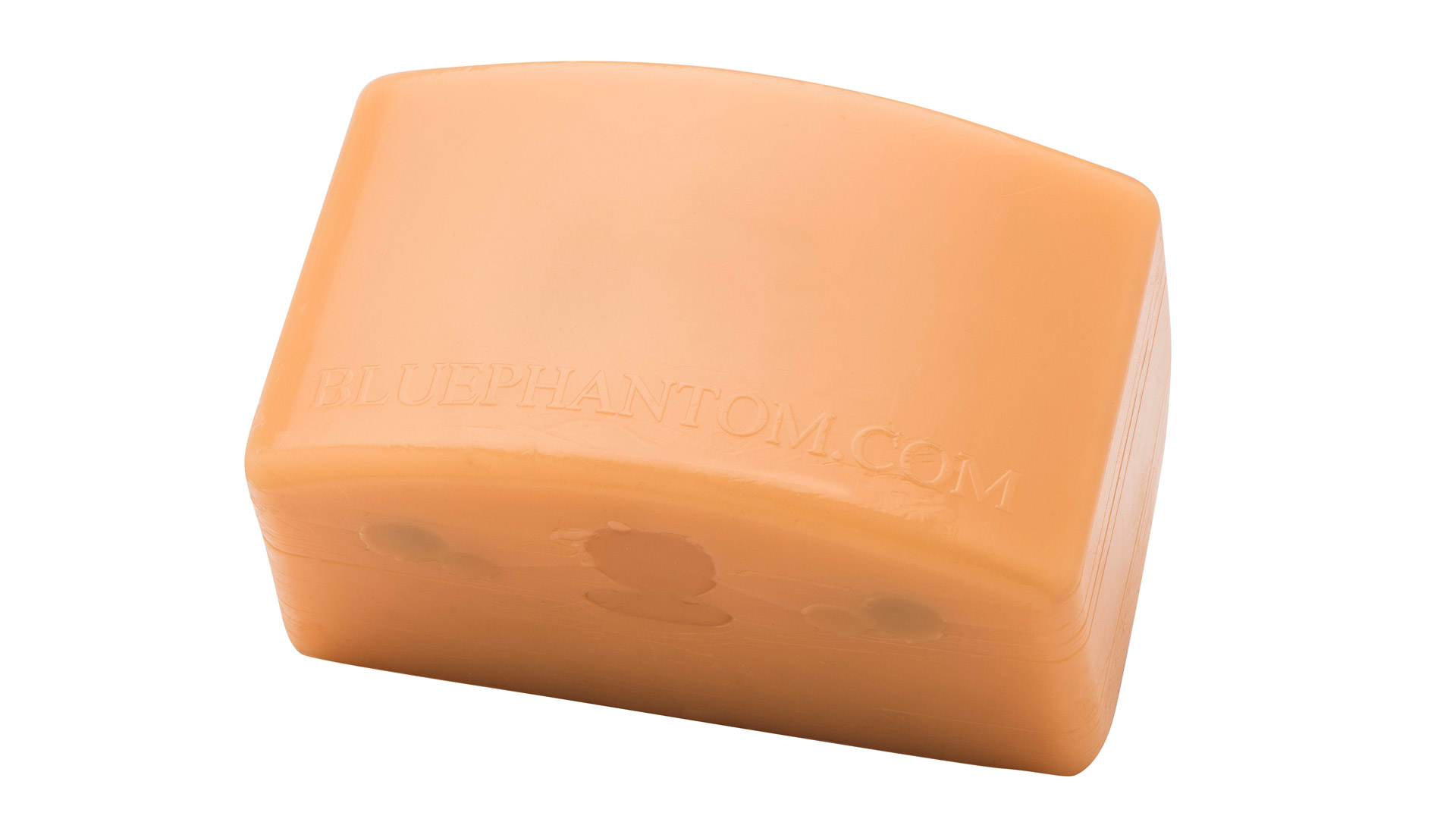
Realistic thyroid anatomy with multiple sonographically distinct lesions
Supports ultrasound-guided fine needle biopsy and diagnostic scanning
Self-healing tissue enables repeated needle insertions without degrading image quality
Includes plastic storage case and preconfigured training pathologies
Includes left and right thyroid lobes, isthmus, multinodular goiter, trachea, esophagus, carotid arteries, and jugular veins—ideal for scanning and biopsy targeting.
Train on a variety of thyroid pathologies with masses ranging in size and echogenicity, including hypoechoic, echogenic, echolucent with echogenic rim, and more.
Build procedural confidence in using ultrasound to locate, target, and biopsy focal lesions with 18–21 gauge needles.
Perform hundreds of needle insertions while maintaining consistent sonographic image quality and model integrity.
Inject fluid to confirm needle placement under ultrasound. Model auto-expels fluid to streamline repeated use.
Excellent for a wide range of learners and clinical specialties including endocrinology, radiology, ENT, and ultrasound education.
Anatomy:
Dimensions:
Included Accessories:
Standard Coverage: 1-Year Manufacturer’s Warranty included
The Blue Phantom Thyroid Biopsy Ultrasound Training Model provides a realistic, repeatable platform for learning and perfecting ultrasound-guided fine needle biopsy (FNB) of the thyroid gland. Constructed using SimulexUS™ self-healing tissue, this model closely mimics the acoustic and tactile properties of real human tissue—making it an essential tool for procedural training, simulation programs, and clinical skills refinement.
Designed for use with any ultrasound imaging system (7.5–15 MHz linear transducer recommended), this training model includes detailed thyroid and surrounding neck anatomy, such as the left and right thyroid lobes, isthmus, trachea, esophagus, carotid arteries, and internal jugular veins. Multiple embedded masses and lesions, including a multinodular goiter and both simple and complex nodules, enable users to develop diagnostic scanning skills and procedural confidence.
Each lesion offers distinct sonographic characteristics—from hypoechoic to echogenic, and echolucent with echogenic or hyperechoic rims—creating a lifelike training environment for FNB targeting and needle visualization. Users can inject fluid to confirm needle tip placement, and the model automatically expels fluid to allow repeated practice without requiring resets.
Whether you’re introducing new learners to neck ultrasound or refining biopsy techniques for experienced providers, this model delivers value through its durability, realism, and cost-effective training experience.