


$10,732.37
Enhance clinical confidence in scrotal ultrasonography with hands-on, pathology-rich ultrasound simulation
Develop diagnostic accuracy through side-by-side comparison of normal and abnormal scrotal anatomy
Practice probe positioning, image acquisition, and measurement using your own ultrasound system
Reinforce learning with true-to-life tissue response and imaging in a risk-free environment
Supports training across emergency, fertility, and urology-based ultrasound education
Includes privacy skirt and soft case for simulation or bedside teaching settings
Learners can scan a healthy testicle and epididymis alongside a pathologic counterpart to reinforce comparative analysis and improve diagnostic precision.
Supports the development of core ultrasound competencies: probe insertion, orientation, scanning planes, and system optimization—critical skills for confident clinical practice.
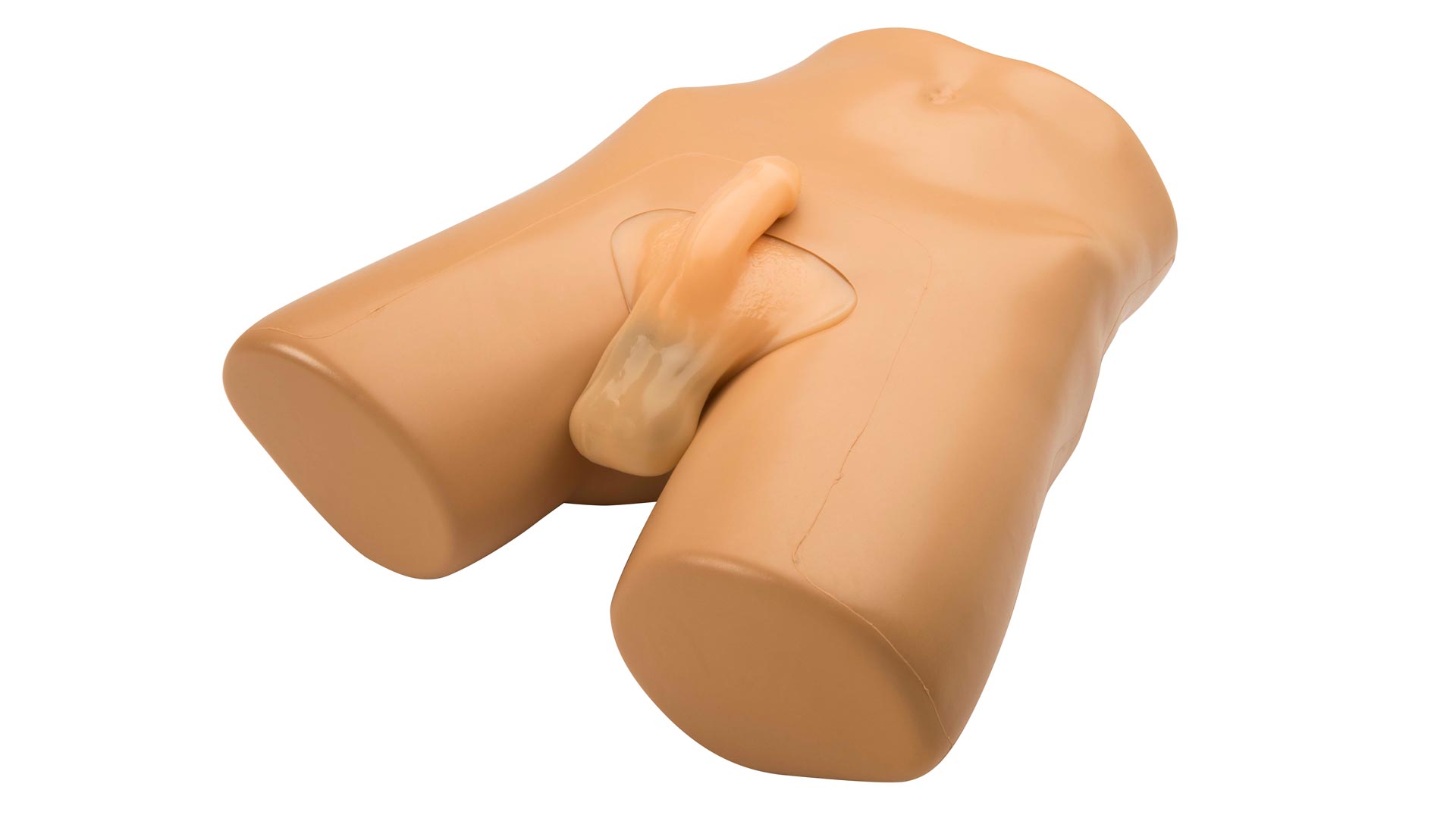
Includes external male pelvic structures—penis, scrotum, and lower pelvis—for an authentic probe positioning experience during training.
Fully imageable structures include both left and right testicles, epididymal head, body, and tail, and pathologies such as intratesticular masses, hydrocele, and an epididymal mass.
Train learners in the real environments they’ll work in. Compatible with any standard ultrasound machine and 2D transducer setup.
Self-healing Simulex™ tissue holds up to repeated scanning without the need for replacement parts. Easy to clean and store.
Anatomy:
Dimensions:
Included Accessories:
Standard Coverage: 1-Year Manufacturer’s Warranty included
The Scrotal Ultrasound Training Model by Blue Phantom is built to support learners in mastering one of the more technically nuanced ultrasound exams: scrotal ultrasonography. Designed for real-time ultrasound use with your own equipment, this model delivers an exceptionally lifelike experience, complete with normal and pathological anatomy for comprehensive training.
Unlike computer simulations or limited static phantoms, this hands-on task trainer mimics the tactile and imaging characteristics of a real patient exam, empowering learners to refine their skills in patient positioning, transducer manipulation, and sonographic interpretation.
Learners can build foundational knowledge by scanning normal anatomy—then immediately reinforce those skills by identifying key abnormalities like intratesticular masses, hydrocele, and epididymal masses. The side-by-side presentation of healthy and pathological anatomy makes it an ideal tool for teaching differential imaging and comparative anatomy.
Whether you’re instructing medical students, training urology fellows, or preparing emergency medicine clinicians, this model offers a risk-free, repeatable platform for teaching the full process of scrotal ultrasound—from setup to interpretation.