$16,188.12
Practice advanced cervical and lumbar spinal procedures with precise anatomical detail and ultrasound imaging performance
Includes cervical (C2–C7) and lumbar (L1–L5) spine anatomy
Enables ultrasound-guided cervical and lumbar epidural training
Realistic CSF flow and adjustable pressure simulation
Durable, self-healing Simulex™ tissue for extended use
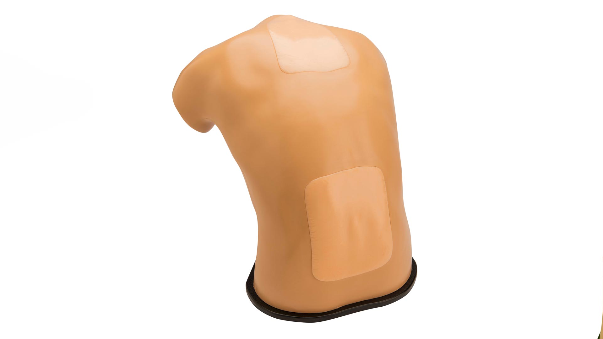
Train on both cervical (C2–C7) and lumbar (L1–L5) procedures using one integrated model for advanced spinal access practice.
Replicates key spinal structures, including ligamentum flavum, epidural and subarachnoid spaces, and dura mater, for authentic tactile response.
Delivers exceptional imaging detail, replicating real patient tissue acoustics for confident needle guidance under ultrasound.
Provides immediate visual and tactile feedback, reinforcing safe and accurate technique.
Simulex™ construction withstands 1,000+ punctures for long-lasting reliability and reduced replacement costs.
Designed as part of Blue Phantom’s modular spinal training platform—interchangeable with lumbar and thoracic inserts.
Anatomy:
• Cervical spine C2–C7
• Lumbar spine L1–L5
• Ligamentum flavum
• Epidural space
• Dura and subarachnoid membrane with CSF
Dimensions:
• Size: 17″ L × 11″ W × 17″ H
• Weight: 33 lb.
Accessories:
• Soft storage case for transport and storage
1-Year Manufacturer’s Warranty included
Usage Requirements: Use only with Blue Phantom ultrasound refill fluid (one bottle included) and sharp, unbent 18–21 gauge needles for optimal performance and warranty coverage.
The Blue Phantom® Lumbar Puncture and Cervical Epidural Ultrasound Training Model provides advanced procedural training for both lumbar puncture and cervical epidural techniques. This dual-region simulator replicates the intricate anatomy and needle paths required for high-risk spinal access procedures.
Learners experience realistic tactile feedback as the needle passes through ligamentum flavum and enters the epidural or subarachnoid spaces, with simulated cerebral spinal fluid (CSF) flow for confirmation of proper placement. The cervical insert offers challenging anatomy that closely mirrors live patient conditions, ideal for training in pain management and regional anesthesia.
Compatible with all ultrasound systems, the model produces high-resolution images across both cervical and lumbar regions, supporting confidence-building in visualization and depth control.