Elevate Healthcare Blog
Gynecological Ultrasound: An Essential Tool for Women’s Health
Jul 15, 2024

The history and evolution of gynecologic ultrasound
Medical ultrasound owes a debt to scientific advances from the 1800s and 1900s like Doppler, radar and sonar. Clinicians saw the potential for diagnostic imaging, and simple models began to emerge in the 1940s and 50s. An early version developed in Minnesota could detect breast tumors. In 1958, a compound contact scanner imaged ascites from ovarian cancer. That same year, a Japanese A scanner helped doctors diagnose cervical cancer.
By 1966, both A-mode (single image) and B-mode (2D) equipment were in common use. By 1972, ovarian follicles could be imaged with static B-mode ultrasound. The technology continued to improve throughout the 70s, with better resolution and diagnostics as well as guidance for invasive procedures.
By the 80s, every system had color Doppler. Mid-decade, the real-time transvaginal probe was introduced; by the 90s it offered both color and harmonic images. And thanks to a French doctor who published stunning first trimester 3D images, 3D/4D imaging became in demand. Today, women’s health ultrasound has expanded to include real time imaging, color and power Doppler, transvaginal sonography, 3D/4D imaging and cell phone-sized handheld devices.
Common gynecological conditions and ultrasound’s role in diagnosis and management
For breast cancer screening, as many as 1 out of 10 American women will need a follow-up ultrasound after a mammogram. In that population, supplemental ultrasound screening shows greater sensitivity and specificity for detecting benign lesions and breast cancer, especially in dense breasts and young women.
Ultrasound can help identify both symptomatic and asymptomatic problems that could progress. For example:
- To evaluate uterine fibroids or ovarian cysts, or determine causes of pelvic pain, abnormal bleeding or other menstrual problems.
- As a follow-up to mammogram with unclear results.
Ultrasound offers a safe, non-invasive, and cost-effective imaging option that allows for real-time visualization and accurate diagnoses, particularly in the early detection of gynecological disorders.
Ultrasound’s importance to women’s health
- Gynecological conditions contribute significantly to women’s health burden. Breast cancer is the most common cancer for women in the U.S., while fibroids account for 30% of all hysterectomies in the U.S.
- Ovarian cysts in women who are past menopause are associated with a higher risk of ovarian cancer.
- Endometriosis affects more than 11% of women ages 15-44 and is linked to ovarian and breast cancer.
- Polycystic ovary syndrome affects 5%-10% of women ages 15-44, and is linked to a number of chronic diseases.
Early detection and treatment of these conditions is critical to women’s overall health and wellbeing. And ultrasound can provide valuable information to aid accurate diagnosis and care. Ultrasound imaging can detect subtle abnormalities and distinguish between benign and malignant masses with high confidence. In the hands of skilled clinicians, advanced technology can help ensure improved patient outcomes and nurture proactive preventive care.
Ultrasound training solutions
Quality education equips learners with the necessary technical skills, and fosters critical thinking, clinical reasoning, and the ability to adapt to evolving healthcare landscapes.
Aspiring ultrasound practitioners face a path filled with challenges, intricacies, and continuous learning. Hands-on training and simulation offers a safe environment to learn and practice. Students have the benefit of a learner-centered environment that allows for extended practice without patient discomfort or harm. They receive real-time feedback on a range of topics, from patient positioning to interpersonal skills, allowing them to develop better technique, compared with traditional training models.
How ultrasound simulation fits into education
Simulation sessions can draw from a variety of technologies, creating an interactive learning experience. It offers compelling advantages, including:
- Efficiency: providing more opportunities to practice skills and gain experience in stressful situations in a risk-free environment.
- Education: more opportunities to increase anatomical understanding beyond initial training.
Simulation can also reduce the burden on clinical staff and increase patient safety during the early stages of ultrasound education.
Educational resources and ongoing training for gynecological ultrasound
Continuous professional development is crucial in order to stay up to date with the latest technologies. Healthcare simulation technology innovation can help drive improvements in patient care and find new solutions to a range of conditions.
Those looking to launch a career in gynecological ultrasound would be best served to find an accredited sonography program. A good resource is the Society of Diagnostic Medical Sonography (SDMS). In addition, facilities and instructors must be licensed with the American Registry for Diagnostic Medical Sonography® (ARDMS®).
Elevate Healthcare: your resource for simulation-based ultrasound education
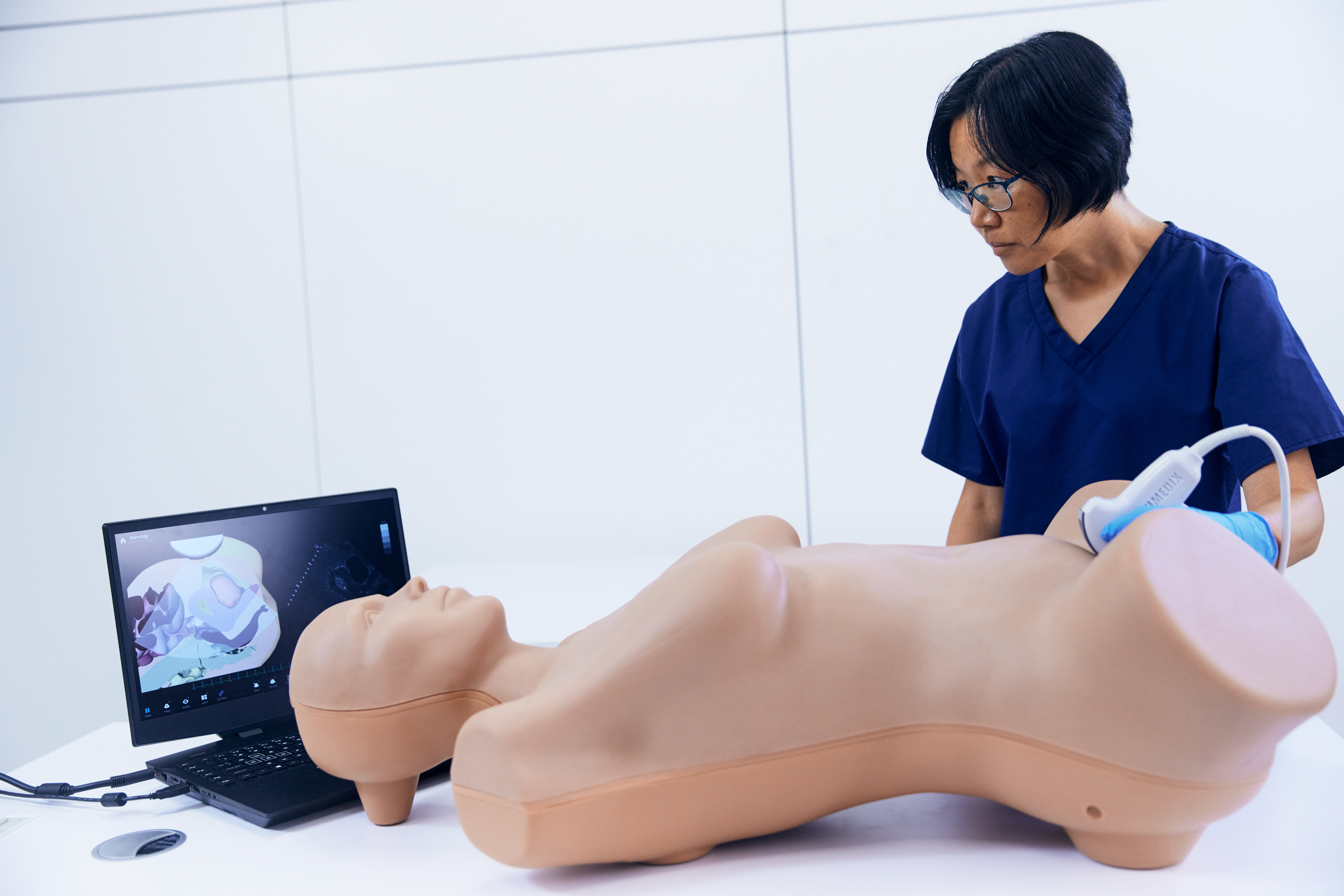
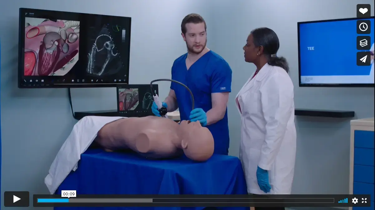
Elevate Healthcare offers a full suite of ultrasound simulation. SimHawk, a unique ultrasound SAAS platform, gives learners an introduction to ultrasound and an opportunity to familiarize themselves with the foundation of ultrasound. From there, students can progress to more advanced training on realistic human models with Vimedix, an ultrasound learning platform featuring both male and female manikins that can simulate thousands of cases.
Next, the Vimedix Ultraportable TEE/ICE Simulator scales down the Vimedix experience to allow trainees to learn probe manipulations in a risk-free environment. Then, they can practice diagnosing and directing treatment on products that mimic human tissue with the Blue Phantom ultrasound trainer.
Elevate Healthcare obstetrics and gynecology solutions
We provide a solution suite designed for teaching and learning the full spectrum of OB-GYN practices, from performing a transvaginal ultrasound exam to delivering a baby with shoulder dystocia.
Our Vimedix includes the complete maternal anatomy and extensive selection of gynecology, including first- and second-trimester pathologies. It offers greater ease of use for both trainers and learners and ultrasound features that can be maximized in different cases. Learn with the curvilinear probe and add on the transvaginal probe for advanced learning.
Medienanfragen
Raven Bouie
Spezialist für kreative Dienstleistungen und Kommunikation
Senden Sie eine E-Mail


